A Positron Emission Tomography (PET) scan is a medical imaging technique used to visualise and assess the functioning of various organs and tissues in the body. It involves the administration of a small amount of radioactive material known as radiotracer, which emits positively charged particles called protons. The interaction between the emitted positrons and surrounding electrons produces gamma rays, which are detected by a special camera to create detailed images.
PET scans are commonly used in clinical settings for diagnosing and monitoring various conditions like cancer, neurological disorders and cardiovascular diseases. The radiotracer used in a PET scan is chosen based on the specific organ or tissue being examined. For example, FDG is commonly used to measure glucose metabolism; making it useful for detecting cancer cells that have increased metabolic activity.
One of the significant advantages of PET scans is their ability to detect abnormalities at the molecular level, even before structural changes become apparent. This makes PET scans highly sensitive in identifying diseases in their early stages. Furthermore, PET scans can help differentiate between benign and malignant tumors, determine the extent of disease spread, and evaluate the effectiveness of treatment.
However, PET scans do have some limitations. They can be expensive and may not be readily available in all healthcare facilities. Additionally, the use of radioactive materials carries a small risk of radiation exposure, although the doses used in PET scans are generally considered safe. Special precautions are taken to minimize radiation exposure, particularly for pregnant women and children.
Read more about: Double marker Test
About Test
About PET Scan Test
When & Why
When & Why
PET scans are performed in various situations and for different reasons, depending on the specific medical condition being evaluated.
Here are some common scenarios in which a PET scan may be done:
- Cancer diagnosis: PET scans are frequently used to detect and evaluate cancerous tumors. They can help identify the location, size, and spread of tumors throughout the body. PET scans are particularly useful in distinguishing between benign and malignant tumors and determining if cancer has metastasized to other organs.
- Treatment Planning and monitoring: PET scans are employed to assess the effectiveness of cancer treatments, such as chemotherapy or radiation therapy. By repeating the scan after treatment, doctors can observe changes in tumor activity and determine if the treatment is shrinking or eliminating the cancerous cells.
- Assessment of heart conditions: PET scans can assess the blood flow to the heart muscle and identify areas with reduced blood supply or damaged tissue. This information aids in diagnosing coronary artery disease, determining the extent of damage from a heart attack, and evaluating the need for further interventions like bypass surgery or angioplasty.

- Brain Disorder: PET scans help in the diagnosis and management of various neurological disorders, including Alzheimer’s disease, Parkinson’s disease, epilepsy, and brain tumors. These scans can visualize abnormal patterns of brain activity and provide valuable information about brain function and metabolism.
- Other conditions: PET scans are also used to evaluate conditions such as infections, inflammation, and certain psychiatric disorders. For example, in cases of suspected infection, a PET scan can help locate the source of infection or determine if it has spread to other areas of the body.
Note: PET scans are often used in conjunction with other imaging techniques, such as CT (computed tomography) or MRI (magnetic resonance imaging), to provide a more comprehensive evaluation. The combined information from multiple imaging modalities helps healthcare professionals make accurate diagnoses and develop appropriate treatment plans for patients.
Test Procedure
Test Procedure
The procedure for a PET scan typically involves the following steps:
- Preparation: Before the scan, you may be instructed to follow certain preparations, such as fasting for a few hours or avoiding certain medications. This is important to ensure accurate results.
- Radiotracer administration: A small amount of radioactive material, called a radiotracer, is administered to you. This can be done through injection, inhalation, or ingestion, depending on the specific radiotracer and the organ or tissue being examined. The radiotracer is designed to accumulate in the area of interest and emit positrons.
- Uptake time: After receiving the radiotracer, you will typically wait for a specific period called the uptake time. This allows the radiotracer to distribute and accumulate in the target area. The duration of the uptake time can vary, ranging from a few minutes to an hour, depending on the radiotracer and the purpose of the scan.
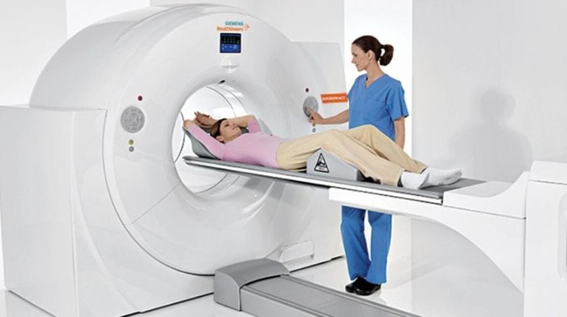
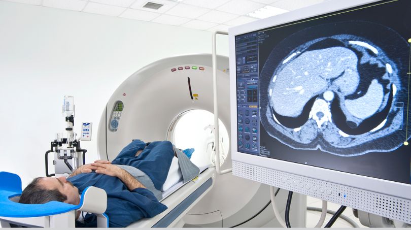
- Imaging: Once the uptake time is complete, you will be positioned on a scanning table. The PET scanner consists of a large circular device with a hole in the center. It may resemble a large doughnut or a cylindrical tube. The table will move slowly into the scanner.

- Scanning: During the scan, you will need to lie still and avoid any movement to ensure clear images. The PET scanner detects the gamma rays emitted by the radiotracer as it decays. It rotates around your body, capturing multiple images from different angles. The scanner does not touch you, and the procedure is painless.
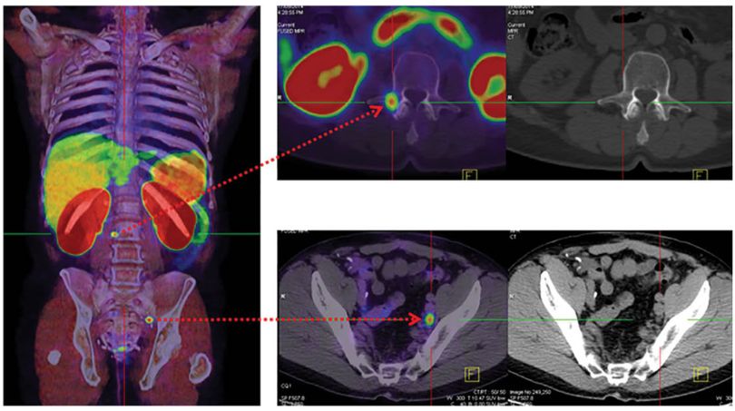
- Additional Imaging: In some cases, a PET scan may be combined with a CT scan or an MRI scan. This is known as a PET-CT or PET-MRI fusion scan. The combination of PET and anatomical imaging helps correlate the functional information provided by PET with the structural details from CT or MRI.
- Post-scan instructions: After the scan, you may be allowed to resume normal activities unless otherwise specified by your healthcare provider. It is essential to drink plenty of fluids to help flush out the radioactive material from your body.
Note: The duration of the scan can vary depending on the area being examined and the specific protocol. In general, a PET scan can take anywhere from 30 minutes to 2 hours to complete.
Normal Range
Normal Range
PET scan provide information about the metabolic activity and function of tissues and organs, allowing healthcare professional’s to assess abnormalities or changes in comparison to normal patterns.
During a PET scan, areas of increased or decreased metabolic activity are highlighted, these area are generally evaluated by comparing them to the normal tissue cells.
Note: Generally an SUV(Standardised uptake value) of 2.5 or higher is considered to be an indication of malignant tissue.
You may like to know about: Troponin Test
Results & Reports
Results & Reports
PET scan results and reports provide a valuable information about the metabolic activity and functioning of tissues and organs. However, some common details that are included in a PET report are:
- Summary of Clinical information
- Radiotracer details
- Imaging findings
- Comparison with other imaging
- Interpretation
- Conclusion and recommendation

How To Book
How To Book
How to book a PET scan test:
- If your healthcare provider determines that a PET scan is necessary, they will provide you with a referral or prescription for the test.
- Research and identify medical facilities in your area that offer PET scan services. You can search online, consult directories, or ask for recommendations from your healthcare provider or friends/family.
- Contact them directly to inquire about the availability of PET scan services and book an appointment. The facility’s contact information, such as phone number or email address, can usually be found on their website or through online search results.
- Once your appointment is scheduled, make sure to receive confirmation of the booking, including the date, time, and any specific instructions for the day of the test. Clarify any doubts or questions you may have, such as what to wear, whether you need someone to accompany you, and if there are any fees or insurance considerations.